Biology

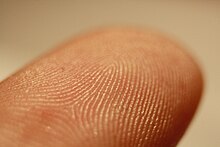
Fingerprints are impressions left on surfaces by the friction ridges on the finger of a human.[1] The matching of two fingerprints is among the most widely used and most reliable biometric techniques. Fingerprint matching considers only the obvious features of a fingerprint.[2]
The composition of fingerprints consists of water (95%-99%), as well as organic and inorganic constituents.[3] The organic component is made up of amino acids, proteins, glucose, lactase, urea, pyruvate, fatty acids and sterols.[3] Inorganic ions such as chloride, sodium, potassium and iron are also present. [3] Other contaminants such as oils found in cosmetics, drugs and their metabolites and food residues may be found in fingerprint residues. [4]
A friction ridge is a raised portion of the epidermis on the digits (fingers and toes), the palm of the hand or the sole of the foot, consisting of one or more connected ridge units of friction ridge skin.[5] These are sometimes known as "epidermal ridges" which are caused by the underlying interface between the dermal papillae of the dermis and the interpapillary (rete) pegs of the epidermis. These unique features are formed at around the 15th week of fetal development and remain with us until after death when decomposition begins.[5] During the development of the fetus, around the 13th week of a pregnancy, ledge-like formation is formed at the bottom of the epidermis beside the dermis.[5] The cells along these ledges begin to rapidly proliferate.[5] This rapid proliferation forms primary and secondary ridges.[5] Both the primary and secondary ridges act as a template for the outer layer of the skin to form the friction ridges seen on the surface of the skin.
These epidermal ridges serve to amplify vibrations triggered, for example, when fingertips brush across an uneven surface, better transmitting the signals to sensory nerves involved in fine texture perception.[6] These ridges may also assist in gripping rough surfaces and may improve surface contact in wet conditions.[7]
Genetics
Consensus within the scientific community suggests that the dermatoglyphic patterns on fingertips are hereditary.[8] The fingerprint patterns between monozygotic twins have been shown to be very similar, whereas dizygotic twins have considerably less similarity.[8] Significant heritability has been identified for 12 dermatoglyphic characteristics.[9] Current models of dermatoglyphic trait inheritance suggest Mendelian transmission with additional effects from either additive or dominant major genes.[10]
Whereas genes determine the general characteristics of patterns and their type, the presence of environmental factors result in the slight differentiation of each fingerprint. However, the relative influences of genetic and environmental effects on fingerprint patterns are generally unclear. One study has suggested that roughly 5% of the total variability is due to small environmental effects, although this was only performed using total ridge count as a metric.[8] Several models of finger ridge formation mechanisms that lead to the vast diversity of fingerprints have been proposed. One model suggests that a buckling instability in the basal cell layer of the fetal epidermis is responsible for developing epidermal ridges.[11] Additionally, blood vessels and nerves may also serve a role in the formation of ridge configurations.[12] Another model indicates that changes in amniotic fluid surrounding each developing finger within the uterus cause corresponding cells on each fingerprint to grow in different microenvironments.[13] For a given individual, these various factors affect each finger differently preventing two fingerprints from being identical while still retaining similar patterns.
It is important to note that the determination of fingerprint inheritance is made difficult by the vast diversity of phenotypes. Classification of a specific pattern is often subjective (lack of consensus on the most appropriate characteristic to measure quantitatively) which complicates analysis of dermatoglyphic patterns. Several modes of inheritance have been suggested and observed for various fingerprint patterns. Total fingerprint ridge count, a commonly used metric of fingerprint pattern size, has been suggested to have a polygenic mode of inheritance and is influenced by multiple additive genes.[8] This hypothesis has been challenged by other research, however, which indicates that ridge counts on individual fingers are genetically independent and lack evidence to support the existence of additive genes influencing pattern formation.[14] Another mode of fingerprint pattern inheritance suggests that the arch pattern on the thumb and on other fingers are inherited as an autosomal dominant trait.[15] Further research on the arch pattern has suggested that a major gene or multifactorial inheritance is responsible for arch pattern heritability.[16] A separate model for the development of the whorl pattern indicates that a single gene or group of linked genes contributes to its inheritance.[17] Furthermore, inheritance of the whorl pattern does not appear to be symmetric in that the pattern is seemingly randomly distributed among the ten fingers of a given individual.[17] In general, comparison of fingerprint patterns between left and right hands suggests an asymmetry in the effects of genes on fingerprint patterns, although this observation requires further analysis.[18]
In addition to proposed models of inheritance, specific genes have been implicated as factors in fingertip pattern formation (their exact mechanism of influencing patterns is still under research). Multivariate linkage analysis of finger ridge counts on individual fingers revealed linkage to chromosome 5q14.1 specifically for the ring, index, and middle fingers.[19] In mice, variants in the gene EVI1 were correlated with dermatoglyphic patterns.[20] EVI1 expression in humans does not directly influence fingerprint patterns but does affect limb and digit formation which in turn may play a role in influencing fingerprint patterns.[20] Genome-wide association studies found single nucleotide polymorphisms within the gene ADAMTS9-AS2 on 3p14.1, which appeared to have an influence on the whorl pattern on all digits.[21] This gene encodes antisense RNA which may inhibit ADAMTS9, which is expressed in the skin. A model of how genetic variants of ADAMTS9-AS2 directly influence whorl development has not yet been proposed.[21]
In February 2023 a study identified WNT, BMP and EDAR as signaling pathways regulating the formation of primary ridges on fingerprints, with the first two having an opposite relationship established by a Turing reaction-diffusion system.[22][23][24]
Classification systems


Before computerization, manual filing systems were used in large fingerprint repositories.[25] A fingerprint classification system groups fingerprints according to their characteristics and therefore helps in the matching of a fingerprint against a large database of fingerprints. A query fingerprint that needs to be matched can therefore be compared with a subset of fingerprints in an existing database.[26] Early classification systems were based on the general ridge patterns, including the presence or absence of circular patterns, of several or all fingers. This allowed the filing and retrieval of paper records in large collections based on friction ridge patterns alone. The most popular systems used the pattern class of each finger to form a numeric key to assist lookup in a filing system. Fingerprint classification systems included the Roscher System, the Juan Vucetich System and the Henry Classification System. The Roscher System was developed in Germany and implemented in both Germany and Japan. The Vucetich System was developed in Argentina and implemented throughout South America. The Henry Classification System was developed in India and implemented in most English-speaking countries.[25]
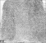
In the Henry Classification System there are three basic fingerprint patterns: loop, whorl, and arch,[27] which constitute 60–65 percent, 30–35 percent, and 5 percent of all fingerprints respectively.[28] There are also more complex classification systems that break down patterns even further, into plain arches or tented arches,[25] and into loops that may be radial or ulnar, depending on the side of the hand toward which the tail points. Ulnar loops start on the pinky-side of the finger, the side closer to the ulna, the lower arm bone. Radial loops start on the thumb-side of the finger, the side closer to the radius. Whorls may also have sub-group classifications including plain whorls, accidental whorls, double loop whorls, peacock's eye, composite, and central pocket loop whorls.[25]
The system used by most experts, although complex, is similar to the Henry Classification System. It consists of five fractions, in which R stands for right, L for left, i for index finger, m for middle finger, t for thumb, r for ring finger and p(pinky) for little finger. The fractions are as follows:
Ri/Rt + Rr/Rm + Lt/Rp + Lm/Li + Lp/Lr
The numbers assigned to each print are based on whether or not they are whorls. A whorl in the first fraction is given a 16, the second an 8, the third a 4, the fourth a 2, and 0 to the last fraction. Arches and loops are assigned values of 0. Lastly, the numbers in the numerator and denominator are added up, using the scheme:
(Ri + Rr + Lt + Lm + Lp)/(Rt + Rm + Rp + Li + Lr)
A 1 is added to both top and bottom, to exclude any possibility of division by zero. For example, if the right ring finger and the left index finger have whorls, the fraction used is:
0/0 + 8/0 + 0/0 + 0/2 + 0/0 + 1/1
The resulting calculation is:
(0 + 8 + 0 + 0 + 0 + 1)/(0 + 0 + 0 + 2 + 0 + 1) = 9/3 = 3
Fingerprint identification

Fingerprint identification, known as dactyloscopy,[29] Ridgeology,[30] or hand print identification, is the process of comparing two instances of friction ridge skin impressions (see Minutiae), from human fingers or toes, or even the palm of the hand or sole of the foot, to determine whether these impressions could have come from the same individual. The flexibility and the randomized formation of the friction ridges on skin means that no two finger or palm prints are ever exactly alike in every detail; even two impressions recorded immediately after each other from the same hand may be slightly different.[31] Fingerprint identification, also referred to as individualization, involves an expert, or an expert computer system operating under threshold scoring rules, determining whether two friction ridge impressions are likely to have originated from the same finger or palm (or toe or sole).
An intentional recording of friction ridges is usually made with black printer's ink rolled across a contrasting white background, typically a white card. Friction ridges can also be recorded digitally, usually on a glass plate, using a technique called Live Scan. A "latent print" is the chance recording of friction ridges deposited on the surface of an object or a wall. Latent prints are invisible to the naked eye, whereas "patent prints" or "plastic prints" are viewable with the unaided eye. Latent prints are often fragmentary and require the use of chemical methods, powder, or alternative light sources in order to be made clear. Sometimes an ordinary bright flashlight will make a latent print visible.
When friction ridges come into contact with a surface that will take a print, material that is on the friction ridges such as perspiration, oil, grease, ink, or blood, will be transferred to the surface. Factors which affect the quality of friction ridge impressions are numerous. Pliability of the skin, deposition pressure, slippage, the material from which the surface is made, the roughness of the surface, and the substance deposited are just some of the various factors which can cause a latent print to appear differently from any known recording of the same friction ridges. Indeed, the conditions surrounding every instance of friction ridge deposition are unique and never duplicated. For these reasons, fingerprint examiners are required to undergo extensive training. The scientific study of fingerprints is called dermatoglyphics.
Limitations and Implications in a Forensic Context
One of the main limitations of friction ridge impression evidence regarding the actual collection would be the surface environment, specifically talking about how porous the surface the impression is on.[32] With non-porous surfaces the residues of the impression will not be absorbed into the material of the surface, but could be smudged by another surface.[32] With porous surfaces, the residues of the impression will be absorbed into the surface.[32] With both resulting in either an impression of no value to examiners or the destruction of the friction ridge impressions.
In order for analysts to correctly positively identify friction ridge patterns and their features depends heavily on the clarity of the impression.[33][5] Therefore the analysis of friction ridges is limited by clarity.[33][5]
In a court context, many have argued that friction ridge identification and Ridgeology should be classified as opinion evidence and not as fact, therefore should be assessed as such.[34] Many have said that friction ridge identification is only legally admissible today because during the time when it was added to the legal system, the admissibility standards were quite low.[35] There are only a limited number of studies that have been conducted to help confirm the science behind this identification process.[5]
Capture and detection
Fingerprinting on Cadavers
The human skin itself, which is a regenerating organ until death, and environmental factors such as lotions and cosmetics, pose challenges when fingerprinting a human. Following the death of a human the skin dries and cools. Fingerprints of dead humans may be obtained during an autopsy.
The collection of fingerprints off of a cadaver can be done in varying ways and depends on the condition of the skin. In the case of cadaver in the later stages of decomposition with dried skin, analysts will boil the skin to recondition/rehydrate it, allowing for moisture to flow back into the skin and resulting in detail friction ridges.[36] Another, method that has been used in brushing a powder, such as baby powder over the tips of the fingers.[37] The powder will ebbed itself into the farrows of the friction ridges allowing for the lifted ridges to be seen.[37]