The umbilical artery is a paired artery (with one for each half of the body) that is found in the abdominal and pelvic regions. In the fetus, it extends into the umbilical cord.
| Umbilical artery | |
|---|---|
 Fetal circulation; the umbilical vein is the large, red vessel at the far left. The umbilical arteries are purple and wrap around the umbilical vein. | |
 Scheme of placental circulation. | |
| Details | |
| Source | Internal iliac artery |
| Branches | Superior vesical artery artery of the ductus deferens |
| Vein | Umbilical vein |
| Identifiers | |
| Latin | arteria umbilicalis |
| MeSH | D014469 |
| TA98 | A12.2.15.020 |
| TA2 | 4316 |
| TE | artery_by_E6.0.1.3.0.0.4 E6.0.1.3.0.0.4 |
| FMA | 18820 |
| Anatomical terminology | |
Structure
Development
The umbilical arteries supply deoxygenated blood from the fetus to the placenta. Although this blood is typically referred to as deoxygenated, this blood is fetal systemic arterial blood and will have the same amount of oxygen and nutrients as blood distributed to the other fetal tissues. There are usually two umbilical arteries present together with one umbilical vein in the umbilical cord. The umbilical arteries surround the urinary bladder and then carry all the deoxygenated blood out of the fetus through the umbilical cord. Inside the placenta, the umbilical arteries connect with each other at a distance of approximately 5 mm from the cord insertion in what is called the Hyrtl anastomosis.[1] Subsequently, they branch into chorionic arteries or intraplacental fetal arteries.[2]
The umbilical arteries are actually the latter of the internal iliac arteries (anterior division of).[3] These supply the hind limbs with blood and nutrients in the fetus.
The umbilical arteries are one of two arteries in the human body, that carry deoxygenated blood, the other being the pulmonary arteries.
The pressure inside the umbilical artery is approximately 50 mmHg.[4] Resistance to blood flow decreases during development as the artery grows wider.[5]
After development
The umbilical artery regresses after birth. A portion obliterates to become the medial umbilical ligament (not to be confused with the median umbilical ligament, a different structure that represents the remnant of the embryonic urachus). A portion remains open as a branch of the anterior division of the internal iliac artery. The umbilical artery is found in the pelvis, and gives rise to the superior vesical arteries. In males, it may also give rise to the artery to the ductus deferens which can be supplied by the inferior vesical artery in some individuals.
Clinical significance
A catheter may be inserted into one of the umbilical arteries of critically ill babies for drawing blood for testing.[6] This is a common procedure in neonatal intensive care, and can often be performed until 2 weeks after birth (when the arteries start to decay too much).[7] The umbilical arteries are typically not suitable for infusions.[6][8]
Additional images
 Model of human embryo, 1.3 mm. long.
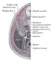 Transverse section of human embryo, eight and a half to nine weeks old.
 Tail end of human embryo, twenty-five to twenty-nine days old.
 Inguinal fossae
 Umbilical artery. Deep dissection. Anterior view.
 Umbilical artery. Deep dissection. Serial cross-section.


See also
References
External links
- Anatomy photo:43:13-0203 at the SUNY Downstate Medical Center - "The Female Pelvis: Branches of Internal Iliac Artery"